
A study conducted by University of Perugia in collaboration with University of Catania and Politecnico di Torino having title Mandible morphing through principal components analysis is focused on the creation of a parametric model of the mandibular bone, with the final aim of setting up a valid tool for the creation of patient-specific morphologies, as an alternative to CT scans, not always available for bone morphology’s definition.
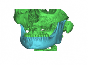
Twenty Computed Tomography scans were used to build the respective 3D CAD models of the mandible. Fifteen of these models were given as an input to a Principal Component Analysis software, and eight ‘principal’ mandible morphologies were produced. The following step was to identify the most efficient landmarks to ‘weight’ these morphologies when building a patient-specific model.
Two further mandible computed tomography scans (a ‘normal’ mandible and a ‘severely resorbed’ one) were used to test the full procedure and to assess its accuracy.
The accuracy of the 3D morphed surface resulted to range between 0.025 and 3.235 mm for the ‘normal’ mandible and between 0.012 and 1.149 mm for the ‘severely resorbed’ one having used eight landmarks to morph a ‘standard’ mandible.
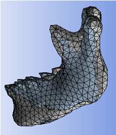
RBF Morph ACT Extension has been used to adapt the baseline mandible mesh onto all the acquired shapes. Having all the variations available for the same mesh topology allowed using the PCA (Principal Component Analysis) method.